|

MISSION of the SAKMAR LABORATORY
at Rockefeller University
Introduction.
The Sakmar Laboratory research program focuses on the molecular mechanism of transmembrane signal transduction by a ubiquitous family of cell-surface receptors called G protein-coupled receptors (GPCRs). GPCRs are serpentine proteins that thread back and forth through the cell membrane seven times. Genes encoding GPCRs make up the largest family in the human genome. GPCRs are responsible for a myriad of signaling processes. For example, specific GPCRs in the retinal detect light and initiate visual phototransduction – color vision depends on the ability of GPCRs sense different colors of light. GPCRs are also responsible for the sense of smell (olfaction) in mammals, and for chemotaxis in lower animals such as flies and worms. A large number of hormones such as adrenaline and glucagon also bind to specific GPCRs to regulate organ physiology in the body. In fact, more than one-quarter of therapeutic drugs targeted to proteins in the human body modulate the activity of GPCRs.
Past Accomplishments.
During his post-doctoral research training at M.I.T. in the mid-1980s, Dr. Sakmar was among the first scientist to study the function of GPCRs using new techniques of molecular biology, in particular heterologous expression and site-directed mutagenesis – a combination of methods where it is possible to introduce amino acid changes at specific sites in the receptor protein and then measure the effect on function. Dr. Sakmar was the first to study mutants of rhodopsin, the receptor for dim-light in the rod cell of the retinal. He was also the first to study the effects of mutations on the ability of GPCRs to couple to another important class of cellular signaling proteins – G proteins.
Dr. Sakmar has made major contributions to understanding the chemical basis for color vision. To detect light, visual GPCRs, sometimes called visual pigments, bind a unique chemical form of vitamin A. Humans have three different visual pigments, which together allow color vision. Dr. Sakmar and his colleagues, in particular the laboratory of Prof. Richard A. Mathies at University of California at Berkeley, identified the specific amino acids in each pigment that “tune” its spectral properties. Using resonance Raman spectroscopy, they also proposed a model for the physical chemistry of spectral tuning.
Dr. Sakmar has also worked on the dynamics of receptor activation – the conformational changes that rapidly occur in the receptor when it absorbs a photon of light or binds to a hormone. It is these conformational changes that carry a signal across the membrane, since the photon or the hormones do not enter the interior of the cell. Dr. Sakmar and colleagues, including Prof. Henry R. Bourne at University of California at San Francisco, proposed the “helix movement model of receptor activation” and put forth the “protonation hypothesis” to explain the regulation of receptor activity. The “helix movement” model is now widely accepted as a key concept in the field of transmembrane signaling.
Computer-generated structural models of GPCRs based on the crystal structure of rhodopsin and other GPCRs have allowed Dr. Sakmar and his colleagues to study specific receptor-drug interactions with the aim of improving the design and synthesis of new drugs with higher potency and fewer side effects. For example, the virus that causes AIDS, HIV, hijacks a GPCR found on lymphocytes, the chemokine receptor 5 (CCR5), to gain entry into the cell. A new class of drugs designed to bind to CCR5 and block HIV cellular entry was recently developed using knowledge of CCR5 structure and biology. Dr. Sakmar and his colleagues, including Prof. John P. Moore at Weill-Cornell Medical College, made key contributions to identifying and optimizing drugs that could binding tightly and specifically to CCR5. The Sakmar Laboratory continues to study the structures of CCR5 and related chemokine receptors and how they become modified by cellular enzymes during biosynthesis. These studies are directly relevant to the molecular pathophysiology of AIDS, and are also relevant to stem cell biology and cancer metastasis, in which chemokine receptors also play key roles.
Interdisciplinary Science.
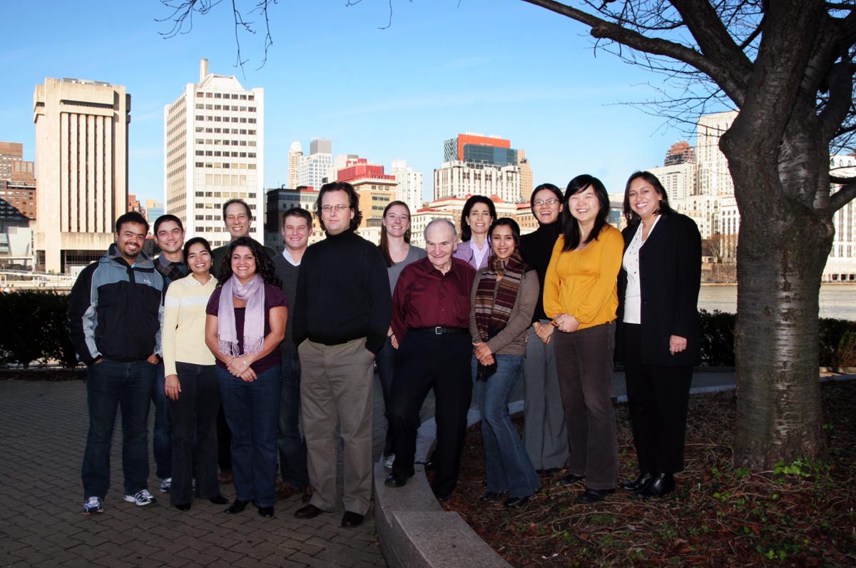
Studies of membrane protein dynamics require expertise is several areas of experimental biology and chemistry. Dr. Sakmar’s laboratory at Rockefeller University currently includes experts in chemical biology, biomolecular spectroscopy, computational chemistry, and molecular biology. Dr. Sakmar and his colleagues have pioneered the development of interdisciplinary experimental approaches to study the molecular mechanism of signal transduction by GPCRs. As noted earlier, with Prof. R. A. Mathies, Dr. Sakmar was the first to study expressed visual pigments by resonance Raman spectroscopy – a laser method that probes the unique vibrations between atoms in the vitamin A chromophore. He also worked with Prof. Friedrich Siebert at Ludwigs-Universität in Freiburg, Germany to provide the first infrared spectra of expressed visual pigments in which the light-dependent movement of specific amino acid groups could be detected. He also worked with Prof. David S. Kliger of University of California at Santa Cruz to study what happens in the first few nanoseconds after light activates a visual pigment. Dr. Sakmar has also worked over many years with Prof. Steven O. Smith at Stony Brook University to develop and exploit solid-state NMR approaches to obtain dynamic structural information about GPCRs in order to highlight the precise movements that occur in the protein’s interior during activation.
Recent Accomplishments.
The interdisciplinary approach of the Sakmar laboratory often results in innovative breakthroughs and new methods and strategies to address important problems in diverse fields. Recent innovations include:
1) The adaptation of an amber codon suppression strategy to introduce unnatural amino acids (UAAs) into engineered GPCRs expressed in mammalian cells in culture, which provides a general tool to label GPCRs with fluorescent probes.
2) The invention of a novel membrane nanostructure, called NABBs (nano-scale apolipoprotein-bound bilayers), to stabilize membrane proteins in a native-like membrane environment.
3) The application of advanced computer methods, including all-atom molecular dynamics simulations and coarse-grain simulations, to the question of GPCR assembly and dynamics in membranes.
4) The discovery that a cellular protein, NucB1, which normally regulates G protein signaling and transport, can inhibit the formation of pathogenic amyloid fibrils by capping smaller growing protofibrils.
Looking Ahead.
For nearly 20 years, the Sakmar Laboratory has been a productive center for innovative studies of GPCRs and related fields, and has also served as a training ground for students at all levels of their scientific training, from post-doctoral fellows to high school students. Working together in the future, members of the Sakmar Laboratory hope to reveal with chemical precision how organisms sense their environment and how cells use chemical signals to communicate. Their studies are directly relevant to drug discovery and to improving therapy for a number of human diseases – AIDS, visual disorders such as retinitis pigmentosa and age-related macular degeneration, and Alzheimer’s disease and other amyloid diseases.
|
 Overview | Highlights | Contact Us | People | Gallery | Research Support